All Categories
History
This section provides an overview for video microscopes as well as their applications and principles. Also, please take a look at the list of 22 video microscope manufacturers and their company rankings. Here are the top-ranked video microscope companies as of July, 2026: 1.Titan Tool Supply, Inc., 2.Lapmaster International, LLC, 3.New York Microscope Company, Inc..
Table of Contents
Categories Related to Video Microscopes

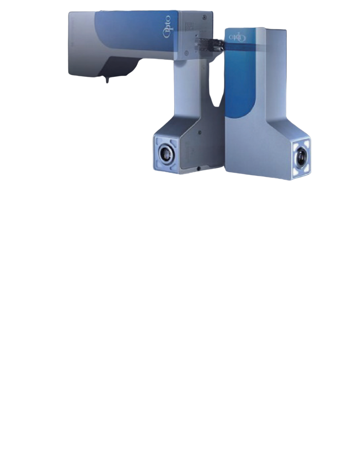
A digital microscope (or video microscope) is an instrument used to magnify an object for observation. However, the term digital microscope generally refers to a microscope equipped with a digital camera and is distinguished from an optical video microscope. Compared to optical video microscopes, digital microscopes have a deeper depth of focus and the ability to measure angles and lengths, which are their main features.
An optical microscope has two lenses, an objective lens, and an eyepiece, while digital microscopes have only an objective lens, and the part corresponding to the eyepiece lens is a digital camera. This can be said to be the most significant difference between an optical microscope and a digital microscope. The digital microscope usually projects the observed object on a monitor.
Several models are available from various manufacturers, with magnifications ranging from several times to several thousand times.
Video microscopes are used not only for magnifying and observing objects but also for evaluations and analyses based on the obtained image data.
They have been introduced in varied fields such as the automotive and aviation industries, the electronic device industries, the medical and cosmetic industries, and the chemical and material industries, and are used in a wide range of applications from research and development to quality assurance.
For example, in failure analysis of electronic components, digital microscopes can be used to inspect the appearance of IC chips, analyze failures of defective products, inspect foreign objects, and analyze their size and shape.
In a digital microscope, an object is magnified by an optical lens (objective lens), and the part that corresponds to the human eye in optical microscopes is a digital camera. The image magnified by the optical lens is detected by the image sensor and the image is displayed on a monitor.
The magnifying power of optical microscopes is expressed as the product of the magnifying power of the objective lens and the eyepiece. In the case of digital microscopes, however, the size of the monitor and the size of the image sensor of the camera affect the magnifying power, which is different from the concept of magnifying power of optical video microscopes. Digital microscope’s magnification is also expressed as the product of the magnification of the objective lens and the magnification of the monitor. The magnifying power of the monitor is calculated by dividing the monitor size by the image sensor size.
In addition to magnification, resolution, or the ability to distinguish details, it is necessary to observe an object in greater detail. If the resolution is not sufficient, the observed image will be blurred and details cannot be observed clearly. In the case of digital microscopes, the resolution of the objective lens, the resolution of the optical lens of the digital camera, the resolution of the image sensor, and the resolution of the monitor all affect the resolution.
It is necessary to select a model that provides optimal magnification and resolution according to the object to be observed and the purpose. To meet user requirements for these advanced resolution processing capabilities, 4K monitor-type images have recently been introduced.
One of the applications of digital microscopes is in dentistry. By taking advantage of the focusing function of digital microscopes, it is possible to observe minute-affected areas that are difficult to detect with the naked eye.
In particular, when performing root canal therapy, which is the complete removal of caries, digital microscopes are used to make it possible for the dentist to remove as much of the affected area as possible.
The use of digital microscopes improves the quality of treatment and reduces the risk of recurrence due to overlooked areas. However, it should be noted that dental treatment using video microscopes is, in principle, not covered by insurance and must be paid out-of-pocket.
Digital microscopes are also used for cosmetic-related treatments and diagnoses, such as cosmetic surgery and scalp checkups. By looking at the skin under microscopic magnification, details such as dryness of the skin and the development of the hairline can be observed.
Clients undergoing cosmetic surgery can also gain a sense of satisfaction from the medical examination by being able to check the condition of their skin and scalp on the screen. It also motivates the client to improve their condition.
Digital microscopes are nowadays often used for detailed analysis of the inside of electronic components and semiconductor ICs down to a few microns, replacing scanning electron microscopes (SEM), which require observation in a vacuum. For this purpose, for practical use, it is necessary to increase magnification and resolution by orders of magnitude from a few millimeters to a few microns during observation.
This operation requires changing the objective lens as in optical video microscopes, but some digital microscopes in recent years have built-in automatic rotation for lens change and automatic focusing function for lens change, making this process almost fully automatic.
In terms of image processing, there are now highly functional types that can combine images with high magnification into a single large image by arranging them vertically and horizontally like tatami mats, and that can process an object into a three-dimensional (3D) image by utilizing the image focus adjustment function.
There are examples where digital microscopes are used to check the wiring of semiconductor ICs and to analyze internal defects in electronic components by combining these functions.
The price of digital (or video) microscopes varies depending on their applications and performance. Digital microscopes with a narrow range of magnification and field of view start at around 10,000 yen, while those used for beauty molding or simple inspection of the scalp are priced at around 50,000 yen, and those used for medical purposes are in the 100,000 yen or more range.
Furthermore, digital microscopes such as those used for product inspection in the manufacturing industry, such as semiconductor manufacturing, require high magnification and micron-level magnification, high-resolution image display, so the price range is generally in the several million yen range.
Low latency screen display and high frame rate are also important for use in surgery and treatment, but digital microscopes with low latency and high frame rate tend to be priced higher. In addition, there are products on the market that allow the magnification of the display to be enlarged by changing the lens. In this case, the image processing capability is also advanced, and the price increases further due to the need for a dedicated monitor and sophisticated control software.
*Including some distributors, etc.
Sort by Features
Sort by Area

Titan Tool Supply was founded in 1952 and operates out of Buffalo, New York. Titan Tool Supply is a supplier of optical instrumentation for research & development, quality control, and other professional industries. Titan Tool Supply’s product lines include borescopes, microscopes, illumination devices, and micro finishing. Titan Tool Supply also offers video systems and magnetics solutions. Titan Tool Supply offers customers a 10-day approval program that allows customers to trial products in their workplace to verify satisfaction before purchase.
LAPMASTER WOLTERS’s roots go back as far as 1804 with the founding of Peter Wolters. Lapmaster was founded in Chicago in 1948 as a manufacture of lapping and polishing machines for the mechanical seal market, Lapmaster has grown to a worldwide solution provider for more than 20 industries like precision optics and advanced materials. Lapmaster serves many industries including automotive, semiconductor, electro optics, and medical devices. Lapmaster also sells many consumables including abrasive powders, lapping compounds diamond lubricants, and composite plates. Products include lapping, polishing, bore honing, buffing, grinding, and special purpose machines.
New York Microscope Company Inc. is based in Hicksville, New York, and is a full-service microscope company. The company serves government, medical, and industrial institutions including the USDA, NASA, Honeywell, and New York University. The company offers 3 varieties of microscopes: compound, stereo, and digital from brands such as Carson, Hitachi, Nikon, Pelican, and others. The company also offers services for repairing, reconditioning, and restoring microscopes. The company can perform calibration tests and fix alignment issues with microscopes.
Penco Precision is a supplier of optical and mechanical inspection equipment established in 1958 in Libertyville, Illinois. The company offers various inspection and optical measurement equipment, including laser micrometers, task light magnifiers, video systems, and force gauges. It also offers gages, lighting products, and torque tools from brands such as Unitron, Mitutoyo, Waldmann, and Starrett. Penco Precision also provides product calibration and repair services to clients in the metal fabrication, medical device manufacturing, automotive, and aerospace industries.

Mitutoyo was founded in 1934 and headquartered in Kawasaki City, Japan. The company is a manufacturer and seller of precision measuring instruments including software, optics, machinery, and electronics. The company is ISO/IEC 17025 accredited for calibration services for measuring instruments. The company’s product lines include Micrometers, Calipers, Calibration Equipment, Vision Measuring Systems, and many others. The company’s coordinate measuring machines and roundness/cylindricity measuring machines are sol in over 60 countries worldwide.

Circuit Imprimé Français (C.I.F) is a France-based manufacturer and supplier of equipment and products used in producing printed circuits boards (PCB) units used in the electronics industry since 1979. Their production line is segmented into equipment for small series & production and laboratory & micro-series equipment including rapid CNC prototyping machines, spray etching machines, PCBs drilling & milling equipment, and direct laser. The company also offers board & stencil cleaning products, compressed air & gas, and furniture such as modular workstations and modular mobile trolleys.

Vision Engineering Ltd, founded in 1958, is a British manufacturer based in Woking, Surrey, specializing in stereo inspection microscopes and non-contact measuring systems. The company's product range encompasses various microscopes, including digital, stereo, and eyepiece-less stereo models. It also offers 3D digital inspection devices integrated with deep reality viewer (DRV) technology, enabling immersive 3D imaging experiences. Additionally, the company provides non-contact measuring systems, spanning from toolmakers' measuring microscopes to fully-automated computerized numerical control (CNC) video measuring systems. Coordinate measuring machines (CMMs) and bench magnifiers are also available. These products find applications in various industries, such as biomedical, electronics, and medical devices.
Olympus is a manufacturer of advanced image, microscopy, and medical equipment. The company was founded in 1919 and headquartered in Tokyo, Japan. The company’s extensive product portfolio includes cutting-edge cameras, lenses, endoscopes, and scientific instruments that have revolutionized businessess and healthcare worldwide. The company combines state-of-the-art technology with unrivalled expertise, setting buisness standards for quality and performance. Olympus empower professionals and enthusiasts alike to capture, visualize, and analyze the world in new and transformative ways.

Optika Italy, established in 1971 and headquartered in Ponteranica, Italy, is a manufacturer and supplier of microscopes and microscopy accessories. The company offers a range of products, including biological microscopes, stereo microscopes, polarizing microscopes, inverted microscopes, and digital cameras with software. These tools find application across diverse sectors such as education, research, industry, and medicine. All of its processes adhere to the Quality Management System, following the UNI EN ISO 9001 standard.
Established in November 1985, Scalar is a manufacturer of optical and electronic devices. They are known for developing the first video loupe in the world. Since then, they have continuously produced innovative products for the medical, beauty, and industrial sectors both domestically and internationally. Their digital microscope, launched in August 1998, has been utilized by NASA as well as airplane and car manufacturing companies. Their team is multidisciplinary and consists of people having experience in optics, physics, electronic engineering, mechanical engineering, chemistry, and computer science to develop cutting-edge products.
Sugitoh Co Ltd, established in 1906, and based in Fujisawa, Japan, is a manufacturer and supplier of optical instruments. The product portfolio includes a diverse range of optical components like optical lens, prisms, special lens, C-mount metal component, beam splitter and other optical instruments. The company also offers optical parts for CCTV and lens, microscope, loupe, image equipment, test equipment, and glass process. The products are used mainly in the medical industry.

Leica Microsystems is a developer and manufacturer of microscopes and scientific instruments for the analysis of microstructures and nanostructures. The company is a provider in compound and stereo microscopy, digital microscopy, confocal user scanning, and super-resolution microscopy with related imaging systems, electron microscopy, sample preparation, and surgical microscopy used in medical, life science, and industrial applications. The company’s services include service plans, preventative maintenance, remote care, and qualification services for ensuring compliance of Leica equipment to governmental regulations.
Wise Device Inc. is a manufacturer and distributor of microscopy automation products that was established in 2003 in Richmond Hill, Ontario, Canada. The company specializes in products for scientific and imaging applications, including high-speed autofocus (ATF) sensors, infrared imaging systems, and precision focus automation (PTF) sensors. It also offers modular microscopy system (MMS) parts, such as illuminators and Z-axis actuators. The company’s products are commonly used in life sciences, photonics, and machine vision.
Saito Kougaku Co. Ltd., established in 2001 and headquartered in Kanagawa, Japan, is a microscope manufacturer and supplier. The company's products include continuous variable magnification systems that integrate with computers, a microscope for full high-definition optical x4 zoom type suitable for use with HD monitors, and a range of accessories that include carry cases, lighting tools, and equipment stands. It also provides a range of software suites to operate its products. These products are used for scientific research in laboratories by all industries, including medical, food, and life sciences.
Lasertec Corporation, founded in 1960 and headquartered in Yokohama, Japan, is a manufacturer and supplier of semiconductor-related systems, FPD-related systems, and laser microscopes. The company's product portfolio includes mask inspection systems, mask edge inspection systems, FPD photomask inspection systems, laser microscopes, and lithium-ion batteries. These products find applications in various sectors, including materials science, the flat panel display industry, semiconductor manufacturing, biomedical and life sciences, as well as research and development. The company is ISO 45001 and ISO 9001 certified, with offices in the USA, China, Singapore, Taiwan, and South Korea for global outreach.
Kurz Ersa has been a manufacturer of machines since 1779 and is based in Kreuzwertheim, Germany. The company operates across diversified business areas, encompassing Electronics Production Equipment, Casting Solutions, Particle Foam Processing, Automation, and Additive Manufacturing. In the realm of Electronics Production Equipment, they offer soldering machines and tools, the Casting Solutions division specializes in light metal casting techniques, and the Particle Foam Processing provides solutions for molded parts. Automation is another domain in which they provide technological solutions. Their expertise in Additive Manufacturing allows delivering of metal 3D printing solutions.
PCE Instruments UK Ltd., started in 199 and headquartered in Manchester, UK, is a manufacturer and supplier of test instruments, equipment, and tools for weighing, measuring, and control systems. The company offers more than 500 test equipment, including analyzers, inspection cameras, meters, detectors, and sensors, with applications in various fields like data acquisition, electrical engineering, environmental science, building inspection, and food processing. Its manufacturing and development division is ISO 9001 certified, all its test instruments, equipment, and tools are factory calibrated, and the company also provides services for custom test instrument design, installation, and maintenance.

BYC INDUSTRIAL LIMITED, a company headquartered in Changsha, China, is a manufacturer and supplier specializing in the production of optical products. The company's range of optical products includes micro zoom lenses, microscope lights, and Newtonian reflectors. These products are used in various applications, including healthcare, jewelry and gemology, as well as astronomy and astrophysics. The company also offers services including technical support to assist clients with product usage, maintenance, and fast delivery of products to customers.
Lighting Specialties, Inc., established in 1917 and headquartered in Chicago, is a distributor and manufacturer of lighting products. The company offers a diverse product catalog including working station lighting, adjustable arm fluorescent lights, waterproof tubular fluorescent fixtures, magnification X-ray viewers, and incandescent waterproof fluorescent products. These lighting solutions cater to a wide array of industries and clientele, including federal and state governments, major industrial distributors, catalog companies, office furniture dealers, and hospitals.
Ranking as of July 2026
Derivation Method| Rank | Company | Click Share |
|---|---|---|
| 1 | Olympus UK & Ireland |
26.3%
|
| 2 | Leica Microsystems Vertrieb GmbH |
17.4%
|
| 3 | Titan Tool Supply, Inc. |
6.1%
|
| 4 | Lapmaster International, LLC |
5.2%
|
| 5 | New York Microscope Company, Inc. |
5.2%
|
| 6 | SUGITOH Co., Ltd. |
4.7%
|
| 7 | C.I.F. – Circuit Imprimé Français |
4.2%
|
| 8 | WDI WISE DEVICE INC. |
2.8%
|
| 9 | Penco Precision |
2.8%
|
| 10 | OPTIKA S.r.l. |
2.8%
|
Derivation Method
The ranking is calculated based on the click share within the video microscope page as of July 2026. Click share is defined as the total number of clicks for all companies during the period divided by the number of clicks for each company.Number of Employees
Newly Established Company
Company with a History
*Including some distributors, etc.
*Including some distributors, etc.
| Country | Number of Companies | Share (%) |
|---|---|---|
|
Japan
|
6 | 35.3% |
|
United States of America
|
4 | 23.5% |
|
United Kingdom
|
3 | 17.6% |
|
France
|
1 | 5.9% |
|
Italy
|
1 | 5.9% |
|
Germany
|
1 | 5.9% |
|
China
|
1 | 5.9% |
316 products found
316 products

Tamapack Co., Ltd.
980+ people viewing
Last viewed: 20 hours ago
No complicated assembly or operation is required. Anyone can use it by simply assembling it and turning on the power. ■CE210T-A Microscope with LC...

Nakamura Choukou Co., Ltd.
510+ people viewing
Last viewed: 1 day ago

Hirox Co., Ltd.
750+ people viewing
Last viewed: 17 hours ago
It is fully equipped with the viewing, photographing, and measuring functions required of a digital microscope, and is compatible with a lens lineu...

Virtue
2310+ people viewing
Last viewed: 2 hours ago
Back up clinical trials related to blood vessels Contribute to research, medical, beauty, and health industry. The license agreement of the blood ...

Star Micro Co., Ltd.
770+ people viewing
Last viewed: 1 day ago
■ Characteristics ・ We have adopted our own dark view lighting mechanism to observe and diagnose the cord quality. ・ The lens has adopted a 500 -...

Micro Square Co., Ltd.
540+ people viewing
Last viewed: 1 day ago
■High resolution and smooth image movement. Full HD microscope with even higher functionality ・Full high-definition microscope DS-330HD is equippe...

Bethel Co., Ltd.
710+ people viewing
Last viewed: 18 minutes ago
■Features ・Thermophysical property microscope is a device that measures thermal effusivity, which is one of the thermophysical property values. ・...

Micro Davans Co., Ltd.
760+ people viewing
Last viewed: 10 hours ago
It is a digital camera -equipped microscope that realizes high -definition shooting of professional specifications. With this one unit, you can mak...

Saito Kogaku
840+ people viewing
Last viewed: 20 hours ago
overview ・ 1920 × 1080p high resolution ・ 1080p Full Hi -Vision (DVI1.0 compliant video output) ・ Progressive scanning, smooth video with 60fps ・ E...

Ptech Co., Ltd.
610+ people viewing
Last viewed: 1 day ago
■Summary The high-performance microscope ``Blood Blood Marpuis II'' is a microscope that allows you to easily observe blood flow and blood vessel s...
AIC-VISION Co., Ltd.
580+ people viewing
Last viewed: 1 day ago
■High-speed imaging machine vision application [Phantom family] is a machine vision camera that achieves excellent high speed. Supports everything ...

Park Systems Japan Co., Ltd.
700+ people viewing
Last viewed: 15 hours ago
SIMON is specially designed for regular measurement tasks in imaging ellipsometry. The simple user interface and robustness of the fixed-angle ell...

Aprolink Co., Ltd.
440+ people viewing
Last viewed: 1 day ago
■Specifications ・Thermography camera & microscope optical system ・Chip-level (micro) thermal imaging of electronic components ・Ultra high precis...
3 models listed

Hirox Co., Ltd.
400+ people viewing
Last viewed: 14 hours ago
A lens series that connects with the digital microscope RH-2000 and RH-8800.

Kokugo Co., Ltd.
650+ people viewing
Last viewed: 1 day ago
Dino-Lite Premier2 M Polarizer LWD DINOAD4113ZTL

Photonic Instruments Co., Ltd.
810+ people viewing
Last viewed: 9 hours ago
ED-Scope is a device for visualizing surface defects and internal defects in optical glass substrates, thin films, wafers, etc. Normally, in the pr...

Three Imaging Co., Ltd.
490+ people viewing
Last viewed: 11 hours ago
■Features Intelli Viewer is software that performs analysis and measurement using a microscope or a camera connected to a high-magnification lens. ...

Irie Co., Ltd.
400+ people viewing
You won't get tired and can see clearly with full high-definition resolution. Details are clearly visible even at low magnification ■Features ・Se...

Anto
2030+ people viewing
Last viewed: 10 hours ago
The capillaries scope "blood vessel beauty" is a device that can easily observe the blood flow of the capillaries without collecting blood. The ob...

GOKO Imaging Devices Co., Ltd.
1050+ people viewing
Last viewed: 1 hour ago
Handheld Multi-distance scope GOKO EV-6HD ・Handheld microscope for professionals ・Has great clarity for professional use, featuring GOKO's prec...

Hirox Co., Ltd.
910+ people viewing
With the addition of various functions to support the operator and the electric control of the lens stand, anyone can easily operate it, and even r...

Star Micro Co., Ltd.
600+ people viewing
Last viewed: 13 hours ago
■ Characteristics The microscope MS-2802 is a revolutionary microscope that enables both TV monitors and PCs because it has a new system function w...

Micro Davans Co., Ltd.
660+ people viewing
This is a digital microscope AS series that has evolved into high -definition image observations by fusion of High Quality camera and high resoluti...
4 models listed

Cosmo Trading Co., Ltd.
1400+ people viewing
Six functions to meet diverse needs. Based on two confocal optical systems, it is equipped with six functions: differential interference observatio...

Koyo Co., Ltd.
510+ people viewing
Last viewed: 1 day ago
A palm-sized, ultra-lightweight CCD camera microscope. The lens and CCD camera body are integrated, and you can easily observe an enlarged image by...

Arms System Co., Ltd.
540+ people viewing
Last viewed: 4 hours ago
This is a digital microscope set with an 11.6 inch full HD display. You can save still images to USB memory, display electronic lines, perform HDR ...

Saito Kogaku
1010+ people viewing
Last viewed: 1 day ago
Detectation Microscope

Shibuya Optical Co., Ltd.
220+ people viewing
■Small, lightweight, and cost-effective microscope unit ・Equipped with infinity correction optical system + coaxial epi-illumination port ・The co...

Micro Square Co., Ltd.
640+ people viewing
Last viewed: 1 day ago
■Compact body ideal for incorporating into equipment. A portable PC microscope. ・Compact body supports magnification from 1x to 400x without chang...
