All Categories
History
This section provides an overview for fluorescence microscopes as well as their applications and principles. Also, please take a look at the list of 11 fluorescence microscope manufacturers and their company rankings. Here are the top-ranked fluorescence microscope companies as of July, 2026: 1.IOLIGHT LIMITED, 2.Etaluma, Inc., 3.PicoQuant.
Table of Contents
Categories Related to Fluorescence Microscopes
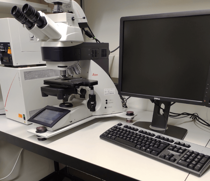
Fluorescence Microscope is a device to observe the fluorescence of fluorescent substances in an object by using laser light, super high-pressure mercury vapor lamp, or xenon lamp as a light source. In ordinary optical microscopes, visible light such as halogen lamps are used as a light source to irradiate an object and observe reflected light and transmitted light.
Fluorescence Microscope is a type of microscope that mainly targets biological tissues and cells labeled with fluorescent substances. Since the resolution of a microscope depends on the wavelength of the light used, Fluorescence Microscope, which uses light with a short wavelength, is characterized by its superior spatial and temporal resolution.
Therefore, highly quantitative information can be obtained. Fluorescence Microscope is becoming more and more important as its functions are becoming more sophisticated as confocal laser microscopy and multiphoton microscopy.
Fluorescence Microscope is mainly used for bio-imaging. The specific targets are cells and tissues, which can be observed while alive. The following techniques are used in combination to label objects with fluorescence:
These technologies enable us to observe the localization of target proteins and expressed genes. In addition, drugs and proteins that emit fluorescence in response to specific substances have been developed, making it possible to visualize neural activity and intracellular dynamics of substances.
In recent years, the advent of CRISPR technology has made the creation of genetically modified organisms much easier, and the scope of its application is rapidly expanding.
Fluorescence Microscope is an instrument for observing fluorescence. Fluorescence is emitted when a fluorescent substance absorbs specific light as energy (excitation light) and releases the energy again.
Exposure to excitation light causes rapid emission of light. The wavelength of fluorescence is longer than the wavelength of the excitation light, and these wavelengths vary with the fluorescent material. Fluorescence Microscope has a filter unit to observe specific fluorescence, which consists of:
By changing or combining filter units, various fluorescent materials can be observed from the same specimen.
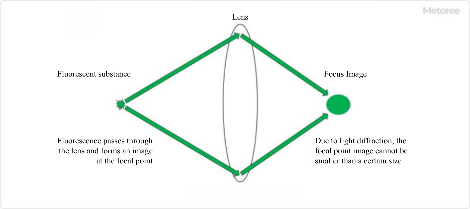
Figure 1. Resolution of fluorescence microscope
The resolution of a microscope refers to "the minimum distance at which two close points can be distinguished from different points. Microscopes use lenses to magnify and observe objects, and in principle, it is possible to increase the magnification infinitely by combining lenses.
However, in the case of optical microscopes, which use light to observe samples, the limit of resolution is approximately half the wavelength of light due to diffraction, which is a characteristic of light. This was considered the theoretical limit of microscope resolution, but a technology was developed that broke through this limit, and the developer was awarded the Nobel Prize in Chemistry in 2014.
The technique is called "super-resolution microscopy. Until the development of super-resolution microscopy, the resolution limit of Fluorescence Microscope was approximately 250 nm, but with super-resolution microscopy, the resolution can be as high as 15-100 nm, which is close to that of electron microscopy. Super-resolution microscopy achieves high resolution by using various techniques to avoid the limiting factors of resolution.
Super-resolution microscopy methods that have dramatically improved resolution and won the Nobel Prize in Chemistry include "PALM" and "STED". PALM and STED have achieved breakthroughs in Fluorescence Microscope resolution by utilizing special optics and special dyes. Super-resolution microscopes using various other technologies have been produced and are being commercialized by various companies.
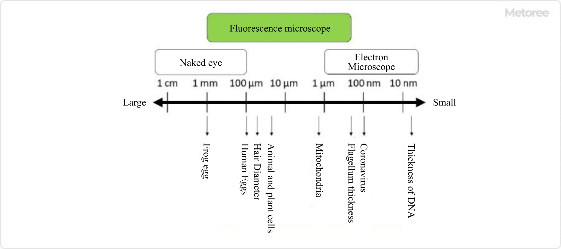
Figure 2. Objects that can be observed with a fluorescence microscope
The advantage of Fluorescence Microscope is that you can observe the behavior of molecules and the structure of cells in detail as visual information. By using the appropriate Fluorescence Microscope for your purpose, you can observe objects with high temporal and spatial resolution.
It is also possible to observe an object using multiple dyes. For example, when two different proteins are labeled with red and green fluorescent substances and observed, a yellow area indicates that these two proteins may exist in the same location in the cell.
A variety of fluorescence materials and Fluorescence Microscopes have been developed for different purposes and applications, and are becoming increasingly important in life science and clinical research.
*Including some distributors, etc.
Sort by Features
Sort by Area

Bruker, founded in 1960 and based in Billerica, Massachusetts, is a manufacturer and distributor of scientific instruments and analytical and diagnostic solutions. The company's product range includes analyzers, microscopes, and imaging solutions, which have applications in fields such as life science research, cell biology, and microbiology. In 1969, the company developed the world's first FT-NMR spectrometer system, enabling broadband proton decoupling. In 1997, it further expanded its capabilities by acquiring the analytical X-ray division of Siemens AG. The company holds ISO 9001 and ISO 13485 certifications, and its products are available for sale worldwide.
Etaluma, Inc., an American company founded in 2009 and based in Carlsbad, California, is a manufacturer and supplier of LS Microscopes (Lumascopes). The company’s product portfolio includes LS820 Microscopes, objective lenses, and phase contrast accessories. These products are primarily used within the life sciences and biomedical research fields, including biomedical research, educational institutions, and clinical and diagnostic laboratories. The company sells its products to various countries including the United Kingdom, China, and Australia. Some of its customers include Pitt Medicine, Stanford Medicine, and Johns Hopkins University.

Olympus is a manufacturer of advanced image, microscopy, and medical equipment. The company was founded in 1919 and headquartered in Tokyo, Japan. The company’s extensive product portfolio includes cutting-edge cameras, lenses, endoscopes, and scientific instruments that have revolutionized businessess and healthcare worldwide. The company combines state-of-the-art technology with unrivalled expertise, setting buisness standards for quality and performance. Olympus empower professionals and enthusiasts alike to capture, visualize, and analyze the world in new and transformative ways.

Leica Microsystems is a developer and manufacturer of microscopes and scientific instruments for the analysis of microstructures and nanostructures. The company is a provider in compound and stereo microscopy, digital microscopy, confocal user scanning, and super-resolution microscopy with related imaging systems, electron microscopy, sample preparation, and surgical microscopy used in medical, life science, and industrial applications. The company’s services include service plans, preventative maintenance, remote care, and qualification services for ensuring compliance of Leica equipment to governmental regulations.
ZEISS Microscopy, established in Jena, Germany, in 1846 is a manufacturer of optics and optoelectronics used in Precision Mechanics, Binoculars, Microscopy, and Eyeglass Lenses. Their product portfolio includes precision optics, such as lenses, mirrors, and prisms, laser mirrors used in laser cutting, and medical devices, optoelectronic devices and Coatings and Thin Films. The company also provides solutions including industrial quality, microscopy research, project simulation, sample testing and product development. The company also offers customer services that include support, custom research projects and customized services.
IOLIGHT LIMITED was founded in 2014 and is located in Whitchurch, United Kingdom is a manufacturer of compact digital microscopes for research purposes and lab incubators. The company provides portable digital microscopes in two models namely 1mm offering higher magnifications and 2mm offering a wider field of view along with portable microscope storage cases, inverted microscopes, and portable fluorescence microscopes, for micro imagining, general biology studies, horticulture, weather forecasting, and bio researching sectors.
Opto GmbH was established in 1980 and is based in Graefelfing, Germany, is a manufacturer of compact imaging modules for industrial and medical applications. The company provides Opto viewer, applications software, OEM imaging modules, multi-fluorescence-microscopes, and, LED illuminations for the test & measurement, life science, research, and, medical markets. They optimize and supplement microscopes in coordination with the manufacturer to enable new set-ups and demanding inspection tasks as well as offer onsite designing services to device developers, machine builders, and OEMs according to the optical requirements of clients.

Dewinter Optical was founded in 1989 and is based in New Delhi, India, is a manufacturer and supplier of microscopes and related image analysis software for various biological and industrial applications. The company provides digital microscope cameras, digital microscopes, fluorescence microscopes, life science microscopes, metallurgical microscopes, and, stereo zoom microscopes in different configurations of monocular, binocular, and trinocular observation tubes, as per customer requirements, for pharmaceutical, biotechnological, metallurgical, and mineralogy industries as well as in semiconductor research, forensic sciences, thin film technology, and QC Laboratories.
ONI was founded in 2016, and is based in Oxford, UK, is a manufacturer and supplier of resolution microscopes for industrial, medical, and research applications. The company provides NimOS & CODI software, EV profiler kit, and, Nanoimager which is a desktop super-resolution microscope, capable of visualizing, tracking, and imaging individual molecules in living cells with 20nm resolution, that have applications in neuroscience, extracellular vesicles, pathogens &immuno oncology, cell phenotyping, and, epigenetic mapping sectors. They work based on single-molecule localization microscopy and offer acquisition, instrument control & basic filtering, powerful analysis, and, storage & collaboration.
PicoQuant, a company founded in 1996 and headquartered in Berlin, Germany, is a manufacturer and supplier of optical and time-resolved measurement instruments. The company’s range of products includes picosecond pulsed drivers, photon counting detectors, and fluorescence spectrometers. These products find applications in various sectors, including materials science, the semiconductor industry, where the instruments are used for quality control and testing of semiconductor materials, and quantum optics and quantum information. The company has global representatives in various countries, including the United States, China, and Canada.

Tokyo Instruments, Inc., established in 1981, and headquartered in Tokyo, Japan, is a manufacturer of opto-electronics products and systems. The company offers a wide range of products, including laser processing machines, aligning systems, photodetectors, analyzers for photoelectron spectroscopy, and spectroscopy array detectors. Also offered are high-speed, high-sensitivity cameras in UV to IR, and photon detectors or counters. The company's product families consist of the Nanofinder series of 3D Laser Raman Microspectroscopy systems and the iDus Workhorse laboratory and OEM CCD platform for low-light spectroscopy.
Ranking as of July 2026
Derivation Method| Rank | Company | Click Share |
|---|---|---|
| 1 | IOLIGHT LIMITED |
16.0%
|
| 2 | Etaluma, Inc. |
11.4%
|
| 3 | Olympus UK & Ireland |
9.4%
|
| 4 | PicoQuant |
9.1%
|
| 5 | Bruker Corporation |
9.1%
|
| 6 | Opto GmbH |
8.7%
|
| 7 | Dewinter Optical Inc. |
8.4%
|
| 8 | ZEISS Microscopy |
8.0%
|
| 9 | ONI. |
7.3%
|
| 10 | Leica Microsystems Vertrieb GmbH |
6.4%
|
Derivation Method
The ranking is calculated based on the click share within the fluorescence microscope page as of July 2026. Click share is defined as the total number of clicks for all companies during the period divided by the number of clicks for each company.Number of Employees
Newly Established Company
Company with a History
*Including some distributors, etc.
*Including some distributors, etc.
| Country | Number of Companies | Share (%) |
|---|---|---|
|
Germany
|
3 | 30.0% |
|
United States of America
|
2 | 20.0% |
|
United Kingdom
|
2 | 20.0% |
| Deutschland | 1 | 10.0% |
|
India
|
1 | 10.0% |
|
Japan
|
1 | 10.0% |
47 products found
47 products

Central Scientific Trading Co., Ltd.
920+ people viewing
Last viewed: 1 day ago
■For those considering purchasing a fluorescence microscope Dual-in-one epifluorescence microscope and optical microscope i4 / Mi-5 Microscope ■Fe...

Brainvision Inc.
1740+ people viewing
Last viewed: 20 hours ago
The THT series is a macro fluorescence microscope that can observe the weak fluorescence emitted from the biological sample at a low magnification ...

Molecular Devices Japan Co., Ltd.
490+ people viewing
Last viewed: 11 hours ago
■ Acquire three-dimensional (3D) images of large organoids and spheroids up to twice as fast The ImageXpress® Confocal HT.ai high-content imaging s...

Brainvision Inc.
800+ people viewing
Last viewed: 26 minutes ago
■Summary A large-diameter tandem lens type low-magnification macro fluorescence microscope developed with the aim of maximally detecting slight cha...